The 26 Review
- What is an X-ray A diagnostic image displaying the bones of the foot
- How Small beams of radiation pass through the foot allowing certain things to show up white, gray or black.
- Why To check for possible signs of injury to the foot and/or ankle
- Recommended For Patients who have sustained trauma or who have unexplained swelling, bruising, and/or pain
Let’s take a closer look…
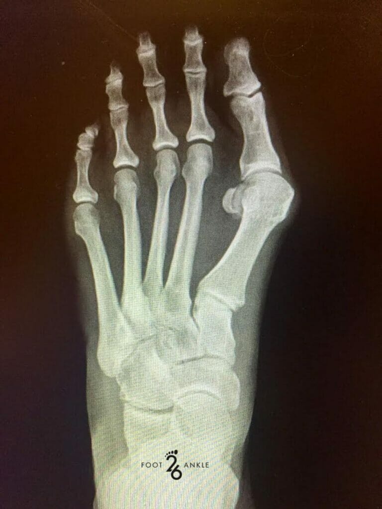
X-rays were discovered for the first time in 1895, making them the oldest medical imaging type used to this day. In conjunction, x-rays are also the most frequently used type of imaging. X-rays or radiographs use a form of radiation called electromagnetic waves and these waves have the ability to capture the inner structures (anatomy) of the body. As mentioned, a foot x-ray creates a black and white image of the inside of the foot. These images display what the bones look like at that given moment. Those bones include the ankle bones (tarsal bones), the toes (phalanges), and the front end of the foot (metatarsal bones). Seeing as different parts of the foot vary in thickness, those areas absorb radiation in different degrees. Calcium found in the body’s bones absorbs more radiation, causing the bones to appear white on x-rays. Soft tissues absorb less radiation, so these areas appear in shades of gray. The air found around the foot will show up in images as black. Healthcare providers utilize foot x-rays to determine the area of injury, extent of injury, to diagnose medical conditions, to locate joint deformities, to watch degenerative conditions, for cysts, bone infections and bone cancer. Patients who have unexplained pain, swelling, bruising, redness and/or tenderness should consider visiting a podiatrist to determine if a foot x-ray is the best diagnostic study. To learn more about foot x-rays or radiographs, continue reading on.
What to Expect
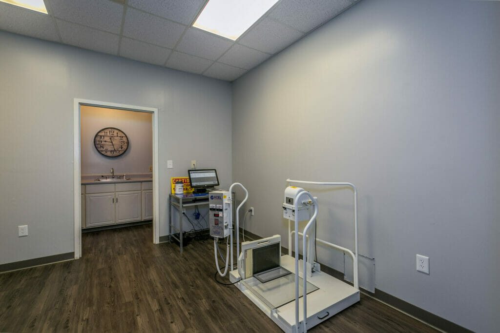
If you or someone you know are receiving a foot x-ray, a radiologic technologist or x-ray technician will likely be performing your/their x-ray. Prior to the x-ray there is not much preparation, other than removing shoes, jewelry or metal objects. Patients who are or may be pregnant should inform the medical professional performing the x-ray. The healthcare provider will determine if the x-ray is truly needed, if yes, precautions will be taken to minimize radiation exposure to the fetus. Before the diagnostic procedure takes place, the technician will explain the full extent of the process, as well as answering any questions.
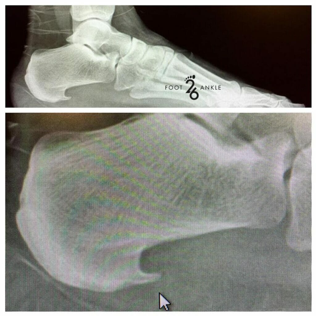
Once shoes are removed in the x-ray room, the technician may provide a lead apron to protect the organs from radiation exposure. To begin, the leg will be placed on the x-ray table and positioning equipment. It is incredibly important to stay as still as possible during the x-ray. Luckily, the entire procedure should take less than a minute. The medical professional may reposition the foot several times to get images from various angles. At the least, 3 separate images are usually taken to get every view. Images will be taken from one side, the front and at a 45 degree angle.
If you are planning to receive an x-ray and experience any pain or discomfort during the procedure, let the technologist know right away. The biggest benefit of an x-ray is that they provide a quick and simple way to view the inside of the foot and diagnose any health conditions and injuries. With foot x-rays, there is a slight amount of radiation exposure and the radiation passes straight through the foot. The benefits of early detection and treatment typically far outweigh the risks.