The 26 Review
- What is a Nail Biopsy A diagnostic procedure to accurately identify infections or ailments
- Types Nail plate biopsy, nail bed biopsy, nail fold biopsy, nail matrix biopsy and longitudinal nail biopsy
- How A sample of toenail and/or tissue is obtained
- Why To guide accurate treatment for a variety of toenail pathologies
- Recommended For Patients that have abnormal appearance, pain, smells, or tenderness located in the nail unit
Let’s take a closer look…
Nail Biopsies are performed to diagnose and treat a large variety of conditions and ailments. This diagnostic study is not simply one technique but rather there are a variety of techniques that can be used. Each style of technique plays a specific role in diagnosis. However in general terms, a nail biopsy takes a sample of tissue of the skin under the nail to check for abnormalities. The different technique biopsies include nail plate, nail bed, nail fold, nail matrix, and longitudinal nail. A nail plate biopsy is somewhat limited in the diagnostic information it can collect, meaning it primarily is used for fungal culture solely. A nail bed biopsy is used to specifically perform skin lesions and rashes isolated under a nail plate, while nail fold biopsies diagnose periungual lesions and rashes. A nail matrix biopsy aids in ruling out melanoma as a potential condition in patients and a longitudinal biopsy is used for large, lateral lesions to determine the underlying cause. The type of biopsy used for a patient will depend on the specific symptoms being felt or seen by a patient. Patients that are experiencing unexplained pain, smells, bruising, redness, and/or tenderness located in the toenail unit should consider speaking with a Podiatrist. Continue reading on to learn more about this diagnostic study.
What to Expect
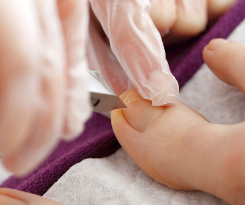
Prior to the biopsy and anesthetic application, the site of the biopsy and surrounding skin will be cleansed thoroughly with an antiseptic cleanser. Following, local anesthesia will then be applied as a nerve block. After anesthetic administration, patients will require roughly a 5-minute waiting period to allow the anesthetic to take full effect. During the waiting period, the toe may be placed in water to soften the nail plate. Once ready, a tourniquet may be applied to the toe to decrease bleeding during the procedure. For certain types of biopsy techniques, the nail will be avulsed (removed) prior to the biopsy. A common avulse technique is with a nail elevator. The nail elevator will be placed under the nail fold to gently loosen the nail plate. Following, the elevator is placed under the distal nail plate and pushed proximally to detach the nail plate from the nail bed and matrix.
As previously mentioned there are a few different biopsy techniques that can be used depending on the circumstance. During a nail plate biopsy a portion of the nail plate is removed with scissors or nail clippers. This type of biopsy doesn’t typically require any anesthetic, however if the nail needs to be partially avulsed prior to the biopsy, a digital block will be administered. To perform a nail bed biopsy the nail is commonly avulsed, which is followed by specimen removal via a punch biopsy tool. With a nail fold biopsy the lesion is removed with a shave/punch tool or a scalpel. A nail elevator tool will also be utilized to access the lesion easier and decrease the risk of damaging the nail matrix. During a nail matrix biopsy the nail is partially or entirely avulsed and the lesion is removed horizontally, rather than longitudinally. This biopsy site is oftentimes sutured for better regrowth of the nail. Finally the longitudinal nail biopsy, this biopsy samples the nail matrix, bed, and fold simultaneously to grant the most diagnostic information. To perform this biopsy, an incision is made in-line with the lateral nail fold between the cuticle and distal interphalangeal joint crease. Another incision is then placed to 3mm medial or lateral to the initial incision. The tissue is then lifted with a skin hook or forceps to remove the tissue all the way down to the bone. This biopsy site is then sutured for optimal healing. This technique is the most invasive with the highest risk for scarring.
Most patients experience throbbing and slight pain 2-3 days after their nail biopsy. If the biopsy site is excessively bleeding, warm to the touch and the pain doesn’t dissipate consider calling your podiatrist.
Pros and Cons
Prior to the procedure, the medical professional will inform the patient of all pre, during and post biopsy expectations. The largest benefit of a nail biopsy is determining the causation of a toenail’s painful symptoms. With most biopsies there are potential risks involved. Some of those risks include bleeding, infection, permanent nail dystrophy, post-operative pain, and the possibility the biopsy doesn’t yield diagnosis. A patient will receive instructions on how to care for the toe after the diagnostic study to prevent possible complications.